December 05, 2005

Bordetella pertussis


Bordetella pertussis

Whooping cough (pertussis) is caused by the bacterium Bordetella pertussis, B. pertussis is a very small Gram-negative aerobic coccobacillus that appears singly or in pairs. Its metabolism is respiratory, never fermentative, and taxonomically, Bordetella is placed among the "Gram-negative Aerobic Rods and Cocci" in Bergey's Manual. Bordetella is not assigned to any family. The bacteria are nutritionally fastidious and are usually cultivated on rich media supplemented with blood. They can be grown in synthetic medium, however, which contains buffer, salts, an amino acid energy source, and growth factors such as nicotinamide (for which there is a strict requirement). Even on blood agar the organism grows slowly and requires 3-6 days to form pinpoint colonies.
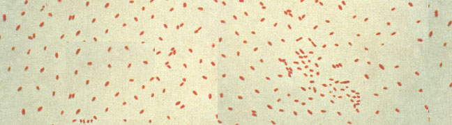
Bordetella pertussis colonizes the cilia of the mammalian respiratory epithelium (Figure 1). Generally, it is thought that B. pertussis does not invade the tissues, but some recent work has shown the bacterium in alveolar macrophages. The bacterium is a pathogen for humans and possibly for higher primates, and no other reservoir is known. Whooping cough is a relatively mild disease in adults but has a significant mortality rate in infants. Until immunization was introduced in the 1930s, whooping cough was one of the most frequent and severe diseases of infants in the United States.
Pathogenesis
The disease pertussis has two stages. The first stage, colonization, is an upper respiratory disease with fever, malaise and coughing, which increases in intensity over about a 10-day period. During this stage the organism can be recovered in large numbers from pharyngeal cultures, and the severity and duration of the disease can be reduced by antimicrobial treatment. Adherence mechanisms of B. pertussis involve a "filamentous hemagglutinin" (FHA), which is a fimbrial-like structure on the bacterial surface, and cell-bound pertussis toxin (PTx). Short range effects of soluble toxins play a role as well in invasion during the colonization stage.

The second or toxemic stage of pertussis follows relatively nonspecific symptoms of the colonizaton stage. It begins gradually with prolonged and paroxysmal coughing that often ends in a characteristic inspiratory gasp (whoop). To hear the characteristic sound of whooping cough click whoop.wav (whoop.wav is copyright of Dr Doug Jenkinson, Nottingham, England. www.whoopingcough.net). During the second stage, B. pertussis can rarely be recovered, and antimicrobial agents have no effect on the progress of the disease. As described below, this stage is mediated by a variety of soluble toxins.

December 01, 2005

Bacillus - General Characterstics

General Characteristics of Bacillus anthracis

· Important pathogen in man and domestic animals for thousands of years
· May have been the 5th plague inflicted on the Egyptians during their negotiations with Moses
· Koch's postulates were developed as a result of his studies on this organism
Capsule
- Composed of poly-(D-glutamic acid), single antigenic type- Nontoxic, serves as an impedin in establishment of infection
- Production enhanced in the presence of Na+-bicarbonate- Capsule gene is plasmid-borne

Spores

- Important in natural history- Form in well aerated cultures (32-35oC), inhibited by high [CO2] (dead carcasses), rare in blood and internal organs- Vegetative phase killed by heat (30 min, 60oC), quickly destroyed in decaying carcasses by enzymatic action and effects of putrefactive bacteria
Colony Characteristics

- Aging colonies: ground glass appearance

Anthrax

· Infects humans and domestic animals
· Usually through contact - Contaminated animal tissue - Wool or hair
· Highly fatal: CFR varies with type of illness

Cutaneous Anthrax

· Spores deposited in abrasion, insect bite
· Germinate, vegetative cells multiply and produce toxin
· Vesicle appears, contains serous fluid which later becomes hemorrhagic and blue-black
· Ruptures, leaving round sharp-edged ulcer with hemorrhagic necrotic tissue
· Ulcer dries, its edges separate from surrounding skin, sloughs off
· Lesion develops fully, results in ulceration, even with appropriate therapy, started early
· 5-20% of untreated patients develop septicemia and generalized infection

Inhalation Anthrax (Woolsorter's Disease)

· Dust particles contaminated with spores are inhaled, deposit in terminal alveoli
· Spores engulfed by macrophages, transported to regional LN
· Germinate, vegetative cells produce toxin
· Extensive necrotic hemorrhage, rapid death frequently results
· Multiple organs involved, CFR about 85% even with Rx

Gastrointestinal Anthrax

· Results from ingestion of contaminated meat
· Organisms or spores penetrate oropharynx/intestinal mucosa
· Deposited in submucosal tissue, multiply and produce toxin
· Usually extends to regional LN, systemic symptoms develop
· CFR about 50%

Anthrax in Domestic Animals

· Major naturally-occurring anthrax areas are tropical, subtropical - India, Pakistan - Africa, South America
· Distribution depends upon conditions allowing sporulation in carcass discharges, vegetative multiplication in soil
· Regions with alkaline soils, high nitrogen level (decaying vegetation) - Alternating periods of rain and drought - Temperatures in excess of 15oC: vegetative multiplication - Resporulation upon drying

Disease in Ruminants

· Sedalia Cattle Trail in Oklahoma, first seeded from dying cattle in the 1800's
· Disease is similar in most ruminants
· Typical presentation is septicemia
· Symptoms: - Sudden onset - High fever, bleeding from body openings - Edema - Peracute death in 1-2h, acute in <24 h
Why is it illegal in some countries to perform post-mortem on antrhax-suspects?
· Diagnosis confirmed by clinical signs, strain/culture from peripheral blood
· Carcasses incinerated on site, buried in quicklime well below ground level

Horses

Symptoms: colic, edematous swellings of the throat, neck, shoulders

Swine, Dogs

Symptoms: pharyngeal swelling, gastroenteritis
· Infection by ingestion of contaminated feed - Raw meat from animals dead of anthrax - Infected meat/bone meal
· Enters from upper part of digestive tract (tonsils)
· Disease manifests as inflamatory edema, tissues of head and neck
· Often become distorted swollen
· Suffocation may follow edema of glottis (tongue)


Virulence Factors and Pathogenesis of Anthrax

"Point of no return:"12 hr antemortem - Guinea pigs inoculated intradermally - Antibody/streptomycin administered before 3 x 10⁸ CFU/ml: course of disease reversed, guinea pigs survived - Rx administered after 3 x 10⁸: death ensued, in spite of substantial reduction in number of bacteria
Findings imply involvement of toxin
Sterile blood from dying guinea pig caused same fatal syndrome in normal guinea pig
Fractionation of plasma revealed three factors:
- I (Edema Factor)
- II (Protective Antigen)
- III (Lethal Factor)

Regulation of EF Activity
EF enzyme activity is calmodulin dependent
No calmodulin in procaryotes
No adenylate cyclase activity in other species of Bacillus

November 30, 2005

Agrobacterium


Agrobacterium tumefaciens (A.t.) is a soil-inhabiting bacterium that causes a disease known as crown gall in many plant species. Roots naturally exude chemicals into the rhizosphere that can be detected by microbes in the soil. A wound can increase the flow of exudates from a plant and specific compounds, such as precursor molecules for lignin (produced by the plant as wound tissue) can stimulate the process of pathogenesis. An organic matrix is created by the bacteria as they attach to the surface of the plant. This matrix facilitates the chance of successful colonization. The cytoplasm of A.t. contains two types of DNA: (1.) a chromosome form and (2.) a smaller, circular piece of DNA called a plasmid. Basically, the plasmid has a short segment (T-DNA) that has genes for hormone production and opine synthesis. Opines are a carbon compound that the bacteria can utilize. This T-DNA is transferred from the bacterial cell through the cell walls of the plant and into the plant cell nucleus. In the nucleus, the T-DNA integrates into the plant chromosome. The cellular processes of the plant treat the T-DNA as it's own and production of the hormones indoleacetic acid and cytokinin begins. Plant cells proliferate undifferentiated tissue, forming a gall. Opines are also produced that are metabolized only by the bacteria. The bacterium may colonize the roots, crown, and other parts of the plant. A.t. has evolved to genetically colonize its host. This is an amazing feat of cross-kingdom genetic engineering by a common soil dwelling bacterium. A computer animation (21 MB mov) illustrates the natural pathogenesis. A second computer animation (16 MB mov) illustrates how this phenomenon is utilized in the lab for Agrobacterium mediated transformation.

Agrobacterium tumefaciens has Gram-negative cell walls.

Flagellar movement of bacteria. A wounded plant cell will beginto synthesize lignin in order to heal the wound.Specific plant lignin precursors are chemotacticallysensed by the pathogen. Flagellarmovement is a series of tumbles and runs counterclockwise upthe gradient and cells have been reported to move as fast as60 um/second.

A. tumefaciens exhibits polar attachment to the plant cell.The production of cellulose fibrils serve to anchor the bacteria to the plantas well as trap other bacteria. Once the concentration of lignin precursorsreaches approximately 10-5 M, the virulence genes of the Ti plasmid are inducedand the T-DNA is processed.

A Ti-plasmid modelis under construction here (Flash)

The model at left shows in an approximate manner how the T-DNA moves into the plant cell and is passedthrough the nuclear pores. The bacterium forms an external pilus (type IV secretion system) for the transfer of T-DNA into the plant cell. The assembly of this secretion system has been studied. A draft animation of this process is available. Type IV Secretion System

November 23, 2005

Part II

Metabolism refers to all the biochemical reactions that occur in a cell or organism. The study of bacterial metabolism focuses on the chemical diversity of substrate oxidations and dissimilation reactions (reactions by which substrate molecules are broken down), which normally function in bacteria to generate energy. Also within the scope of bacterial metabolism is the study of the uptake and utilization of the inorganic or organic compounds required for growth and maintenance of a cellular steady state (assimilation reactions). These respective exergonic (energy-yielding) and endergonic (energy-requiring) reactions are catalyzed within the living bacterial cell by integrated enzyme systems, the end result being self-replication of the cell. The capability of microbial cells to live, function, and replicate in an appropriate chemical milieu (such as a bacterial culture medium) and the chemical changes that result during this transformation constitute the scope of bacterial metabolism.
The bacterial cell is a highly specialized energy transformer. Chemical energy generated by substrate oxidations is conserved by formation of high-energy compounds such as adenosine diphosphate (ADP) and adenosine triphosphate (ATP) or compounds containing the thioester bond

(acetyl ~ SCoA) or succinyl ~ SCoA. ADP and ATP represent adenosine monophosphate (AMP) plus one and two high-energy phosphates (AMP ~ P and AMP ~ P~ P, respectively); the energy is stored in these compounds as high-energy phosphate bonds. In the presence of proper enzyme systems, these compounds can be used as energy sources to synthesize the new complex organic compounds needed by the cell. All living cells must maintain steady-state biochemical reactions for the formation and use of such high-energy compounds.
Kluyver and Donker (1924 to 1926) recognized that bacterial cells, regardless of species, were in many respects similar chemically to all other living cells. For example, these investigators recognized that hydrogen transfer is a common and fundamental feature of all metabolic processes. Bacteria, like mammalian and plant cells, use ATP or the high-energy phosphate bond (~ P) as the primary chemical energy source. Bacteria also require the B-complex vitamins as functional coenzymes for many oxidation-reduction reactions needed for growth and energy transformation. An organism such as Thiobacillus thiooxidans, grown in a medium containing only sulfur and inorganic salts, synthesizes large amounts of thiamine, riboflavine, nicotinic acid, pantothenic acid, pyridoxine, and biotin. Therefore, Kluyver proposed the unity theory of biochemistry (Die Einheit in der Biochemie), which states that all basic enzymatic reactions which support and maintain life processes within cells of organisms, had more similarities than differences. This concept of biochemical unity stimulated many investigators to use bacteria as model systems for studying related eukaryotic, plant and animal biochemical reactions that are essentially "identical" at the molecular level.
From a nutritional, or metabolic, viewpoint, three major physiologic types of bacteria exist: the heterotrophs (or chemoorganotrophs), the autotrophs (or chemolithotrophs), and the photosynthetic bacteria (or phototrophs) (Table 4-1). These are discussed below.
Heterotrophic Metabolism
Heterotrophic bacteria, which include all pathogens, obtain energy from oxidation of organic compounds. Carbohydrates (particularly glucose), lipids, and protein are the most commonly oxidized compounds. Biologic oxidation of these organic compounds by bacteria results in synthesis of ATP as the chemical energy source. This process also permits generation of simpler organic compounds (precursor molecules) needed by the bacteria cell for biosynthetic or assimilatory reactions.

The Krebs cycle intermediate compounds serve as precursor molecules (building blocks) for the energy-requiring biosynthesis of complex organic compounds in bacteria. Degradation reactions that simultaneously produce energy and generate precursor molecules for the biosynthesis of new cellular constituents are called amphibolic.
All heterotrophic bacteria require preformed organic compounds. These carbon- and nitrogen-containing compounds are growth substrates, which are used aerobically or anaerobically to generate reducing equivalents (e.g., reduced nicotinamide adenine dinucleotide; NADH + H+); these reducing equivalents in turn are chemical energy sources for all biologic oxidative and fermentative systems. Heterotrophs are the most commonly studied bacteria; they grow readily in media containing carbohydrates, proteins, or other complex nutrients such as blood. Also, growth media may be enriched by the addition of other naturally occurring compounds such as milk (to study lactic acid bacteria) or hydrocarbons (to study hydrocarbon-oxidizing organisms).

November 16, 2005

THE DIVERSITY OF METABOLISM IN PROCARYOTES

Introduction
A lot of hoopla is made about microbial diversity.  Based on superficial inspection, the bacteria and archea hardly seem diversified. There are but a few basic morphologies, the possibilities of motility and resting cells (spores), and a major differential stain (the Gram stain) to distinguish the procaryotes microscopically. In the eukaryotes, there may be more structural diversity within a single genus of organisms. So what is all the hoopla about? It is about biochemical or metabolic diversity, especially as it relates to energy-generating metabolism and biosynthesis of secondary metabolites. The procaryotes, as a group, conduct all the same types of basic metabolism as eukaryotes, but, in addition, there are several types of energy-generating metabolism among the procaryotes that are non existent in eukaryotic cells or organisms. The diversity of procaryotes is expressed by their great variation in modes of energy production and metabolism.
Even within a procaryotic species, there may be great versatility in metabolism. Consider Escherichia coli. The bacterium can produce energy for growth by fermentation or respiration. It can respire aerobically using O2 as a final electron acceptor, or it can respire under anaerobic conditions, using NO3 or fumarate as a terminal electron acceptor. E. coli can use glucose or lactose as a sole carbon source for growth, with the metabolic ability to transform the sugar into all the necessary amino acids, vitamins and nucleotides that make up cells. A relative of E. coli, Rhodospirillum rubrum, has all the heterotrophic capabilities as E. coli,plus the ability to grow by photoautotrophic, photoheterotrophic or lithotrophic means. It does require one growth factor, however; biotin must be added to its growth media.
Fundamentally, most eukaryotes produce energy (ATP) through alcohol fermentation (e.g. yeast), lactic acid fermentation (e.g. muscle cells, neutrophils), aerobic respiration (e.g. molds, protozoa, animals) or oxygenic photosynthesis (e.g. algae, plants). These modes of energy-generating metabolism exist among procaryotes, in addition to all following types of energy production which are virtually non existent in eukaryotes.
Unique fermentations proceeding through the Embden-Meyerhof pathway
Other fermentation pathways such as the phosphoketolase (heterolactic) and Entner-Doudoroff pathways
Anaerobic respiration: respiration that uses substances other than 02 as a final electron acceptor
Lithotrophy: use of inorganic substances as sources of energy
Photoheterotrophy: use of organic compounds as a carbon source during bacterial photosynthesis
Anoxygenic photosynthesis: photophosphorylation in the absence of O2
Methanogenesis: an ancient type of archaeon metabolism that uses H2 as an energy source and produces methane
Light-driven nonphotosynthetic photophosphorylation: unique archaeon metabolism that converts light energy into chemical energy
In addition, among autotrophic procaryotes, there are three ways to fix CO2, two of which are unknown among eukaryotes, the CODH (acetyl CoA pathway) and the reverse TCA cycle.

November 15, 2005

Bacterial Metabolism


Heterotrophic Metabolism
Heterotrophic metabolism is the biologic oxidation of organic compounds, such as glucose, to yield ATP and simpler organic (or inorganic) compounds, which are needed by the bacterial cell for biosynthetic or assimilatory reactions.
Respiration
Respiration is a type of heterotrophic metabolism that uses oxygen and in which 38 moles of ATP are derived from the oxidation of 1 mole of glucose, yielding 380,000 cal. (An additional 308,000 cal is lost as heat.)
Fermentation
In fermentation, another type of heterotrophic metabolism, an organic compound rather than oxygen is the terminal electron (or hydrogen) acceptor. Less energy is generated from this incomplete form of glucose oxidation, but the process supports anaerobic growth.
Krebs Cycle
The Krebs cycle is the oxidative process in respiration by which pyruvate (via acetyl coenzyme A) is completely decarboxylated to C02. The pathway yields 15 moles of ATP (150,000 calories).
Glyoxylate Cycle
The glyoxylate cycle, which occurs in some bacteria, is a modification of the Krebs cycle. Acetyl coenzyme A is generated directly from oxidation of fatty acids or other lipid compounds.
Electron Transport and Oxidative Phosphorylation
In the final stage of respiration, ATP is formed through a series of electron transfer reactions within the cytoplasmic membrane that drive the oxidative phosphorylation of ADP to ATP. Bacteria use various flavins, cytochrome, and non-heme iron components as well as multiple cytochrome oxidases for this process.
Mitchell or Proton Extrusion Hypothesis
The Mitchell hypothesis explains the energy conservation in all cells on the basis of the selective extrusion of H+ ions across a proton-impermeable membrane, which generates a proton motive force. This energy allows for ATP synthesis both in respiration and photosynthesis.
Bacterial Photosynthesis
Bacterial photosynthesis is a light-dependent, anaerobic mode of metabolism. Carbon dioxide is reduced to glucose, which is used for both biosynthesis and energy production. Depending on the hydrogen source used to reduce CO2, both photolithotrophic and photoorganotrophic reactions exist in bacteria.
Autotrophy
Autotrophy is a unique form of metabolism found only in bacteria. Inorganic compounds are oxidized directly (without using sunlight) to yield energy (e.g., NH3, NO2-, S2, and Fe2+). This metabolic mode also requires energy for CO2 reduction, like photosynthesis, but no lipid-mediated processes are involved. This metabolic mode has also been called chemotrophy, chemoautotrophy, or chemolithotrophy.

Anaerobic Respiration
Anaerobic respiration is another heterotrophic mode of metabolism in which a specific compound other than 02 serves as a terminal electron acceptor. Such acceptor compounds include NO3-, SO42-, fumarate, and even CO2 for methane-producing bacteria.
The Nitrogen Cycle
The nitrogen cycle consists of a recycling process by which organic and inorganic nitrogen compounds are used metabolically and recycled among bacteria, plants, and animals. Important processes, including ammonification, mineralization, nitrification, denitrification, and nitrogen fixation, are carried out primarily by bacteria.

November 09, 2005

Bacterial Lysis

Bacterial Killing
Aims and Objectives
  • To understand the terms complement, opsonin, opsonisation, phagocytosis.

  • To understand the principles of complement activation.

  • How complement is involved in the elimination of bacteria.

  • To understand the types of cell involved in phagocytosis.

  • To understand the process of phagocytosis.

  • To understand the biochemical mechanisms of killing and degradation of bacteria by phagocytes.

  • To understand the clinical features and management of patient's with antibody, complement or neutrophil deficiences.
Complement, Opsonisation and Phagocytosis
Opsonisation is the process of coating micro-organisms with plasma proteins to increase their adherence to phagocytic cells in preparation for phagocytosis. The two main opsonins are IgG antibody and the third component of complement (C3) which bind to the surfaces of micro-organisms. Phagocytic cells have membrane receptors for IgG (Fc piece) and activated C3 (which is called C3b). IgG antibody binds to micro-organisms because the Fab portion recognises bacterial epitopes. C3b binds because during activation of C3 a thiol bond is exposed which permits the formation of ester or amide bonds (covalent binding) with the bacterial surface structures.
C3 is activated by limited proteolysis by enzymes called C3 convertases. There are two C3 convertases, one produced by activation of the classical pathway which is antibody dependent (IgM or IgG), while the alternative pathway can be activated in the absence of antibody. The classical pathway is inactivated when the C1q subcomponent of first component of complement binds to two adjacent IgG molecules or a single IgM molecule. The C1s subcomponent of C1 becomes an active protease and activates C4 and C2 to form the classical pathway C3 convertase which is a complex of activated C4 (C4b) and C2 (C2a) it is designated C4b2a. The alternative pathway is activated when micro-organisms come into contact with body fluids. The pathway is always being activated slowly to generate small amounts of activated C3 (C3b). When C3b binds to a micro-organism activation of the alternative pathway is amplified so that a large amount of the alternative pathway C3 convertase is formed on the surface, followed by deposition of activated C3 (C3b).
Complement can also kill micro-organisms directly. Once C3b has been formed, same binds to the C3 convertase to change it to a C5 convertase which activates C5 to form C5b. Subsequently C6, C7, C8 and C9 bind together with C5b to form a hollow cylinder which is inserted into cell membranes to produce lysis.

Thus complement kills micro-organisms in two ways:
  1. Opsonisation followed by phagocytosis and intracellular killing (indirect).

  2. Assembly of the cytolytic C5b-9 membrane attack complex (direct).
Neutrophils and monocytes/macrophages are the two important phagocytic cells. Neutrophils and monocytes circulate in the blood and migrate into the tissues at the post-capillary venule. Initially they adhere to endothelium and then migrate through intercellular junctions. In the tissues they migrate towards bacteria by means of a process called chemotaxis, which is defined as direct movement along a concentration gradients of chemotactic agents (e.g. C5a leukotriene B4, IL-8, bacterial peptides). Phagocytes recognise their targets by specific sugar residues (e.g. mannose or LPS) but binding is greatly enhanced if the organism is opsonised with IgG and/or C3b. Phagocytes possess Fc and C3b receptors and there is co-operation between these receptors. Thus an organism opsonised with IgG and C3b is more effectively phagocytosed.
Ingestion (phagocytosis) is a localised endocytosis process requiring energy. The plasma membrane envelopes the particle and buds off to form an intracellular vesicle, the phagosome. Following fusion of the phagosome with lysosomal granules the phagolysosome is formed and the bacteria are killed by oxygen-dependent and oxygen-independent process.

Intracellular killing mechanisms:

Oxygen dependent:
  • Hydrogen peroxide

  • Sight oxygen

  • Hydroxyl radical

  • Hypohalite

  • Nitric oxid
Oxygen independent:
  • Lysozyme

  • Lysosomal contents

  • acid hydrolases

  • cationic proteins

  • Lactoferrin

  • Neutral proteases

November 01, 2005

Facts on Bacteria

What are bacteria?
Bacteria are a successful and ancient form of life, quite different from the eukaryotes (which includes the fungi, plants and animals). They are small cells, found in the environment as either individual cells or aggregated together as clumps, and their intracellular structure is far simpler than eukaryotes. Bacteria have a single circular DNA chromosome that is found within the cytoplasm of the cell as they do not have a nucleus. Indeed they lack any of the intracellular organelles so characteristic of eukaryotic cells, such that they do not have the golgi apparatus, endoplasmic reticulum, lysosomes nor mitochondria. However they are generally capable of `free-living' and therefore they possess all the biosynthetic machinery that is needed for this, including 70S ribosomes (as opposed to the larger 80S forms found in eukaryotes) distributed throughout the cytoplasm. The most complex region of the cell is often the cell surface. The cell wall / outer membrane is described below, but in addition some bacteria may secrete a polysaccharide capsule onto their outer surface, some may have flagella which they require for mobility and some may have external projections such as fimbriae and pili which are useful for adherence in their chosen habitat. Although bacteria are generally far simpler than eukaryotic cells, they are extremely efficient within their own little niche - and this may include the ability to cause human infections. Bacteria multiply by binary fission and there is no sexual interaction.
Gram stain appearances of medically important bacteria
As bacteria are so small, they need to be viewed under a microscope using special stains; the stain that is traditionally used for this is called the "Gram stain". In this process, purple dyes are poured over bacteria that have been spread out thinly on a microscope slide and the cell walls of the bacteria (made out of peptidoglycan) take up the colour. If a solvent is then applied to the slide, bacteria which have only got a cell wall still keep their purple colour, but bacteria which have got an extra cell membrane (made out of phospholipid) outside their cell wall quickly lose the purple stain and become colourless; in order to be able to see these bacteria under the microscope a second red stain is then used.
  • Bacteria that manage to keep the original purple dye have only got a cell wall - they are called Gram positive.

  • Bacteria that lose the original purple dye and can therefore take up the second red dye have got both a cell wall and a cell membrane - they are called Gram negative.
A table of the staining characteristics for some common bacteria of medical importance is given below. Note that for cocci, it is not just the shape and colour of the individual bacterial cells that is important, but the way that all these cells group together too. Put simply, round purple balls that look like bunches of grapes under the microscope (i.e. Gram-positive cocci in clusters!) suggests that the bacteria are staphylococci. Most of these bacteria are fairly flexible about the conditions they require for their growth - give them roughly the right temperature and a few simple nutrients and they are happy. A few of them are rather more fussy though and bacteria such as Clostridium and Bacteroides are examples of this; they are called anaerobes which means that they can only grow if there is absolutely no oxygen present.


Why should you bother with all this? There are three reasons:
  • dividing up new information makes it easier to learn - and unfortunately there is no option but to learn the core material in this table.

  • this is the language used by clinicians, especially when they are trying to manage seriously ill patients.

  • knowing this information well makes it easier to understand bacteria, to appreciate the different sorts of infections they cause and the antibiotics that can be used to treat them.
Exceptions to the rule: not all bacteria are shown up by the Gram stain, but there are only a couple of important exceptions for you to remember now!
  • Mycobacterium tuberculosis has a thick waxy coat that stops the bacterial cells taking up the Gram stain; if these bacteria are suspected, the specific Ziehl Neelson stain must be used.
  • A small group of organisms, such as Chlamydia and Mycoplasma, do not have conventional cell walls at all and specialised techniques are often required to diagnose infections caused by these bacteria.

How do you get information about bacteria in clinical practice?
A number of different samples can be sent to the diagnostic laboratory for microbiological analysis including fluids (such as blood, urine or cerebrospinal fluid [CSF]), pieces of tissue or swabs taken from infected lesions. For specimens such as CSF that would normally be sterile, microscopy can be very useful as the presence of any bacteria is always abnormal. However, for the great majority of specimens, the sample will have to be spread out onto culture plates to grow the bacteria, to see if there are any in the sample that might be the cause of the infection - this will generally take at least 24 hours. If there are any suspect bacteria there, they will probably need to be identified further and also checked out to ensure that they are not resistant to the effect of antibiotics - at least another 24 hours. In situations where it is not possible to grow bacteria, it may be possible to diagnose infection based upon a person's antibody responses - but this is not usually a rapid method either! So that in many cases, you must make initial decisions about antibiotic treatment based upon a sound knowledge of bacteriology - what is the most likely or most important bacterial cause of this infection and what is the most appropriate antibiotic for treatment? This will usually be backed up by laboratory investigation to confirm your diagnosis (or not!) and help you refine your future management of the patient. To practice clinical medicine effectively you must have a good knowledge of bacteriology, some idea about the service provided by diagnostic laboratories and the ability to interpret the reports that are issued.
In the table of Gram stains above, the bacteria were grouped together and listed by genus name (plural = genera). This is a name given to a collection of bacteria that share many fundamental, major, obvious characteristics. However, by examining bacteria more closely, perhaps by looking at some of their biochemical capabilities for example, it is possible to divide them up further into individual species. This is very often of great clinical significance. An example of how this works in clinical practice is given below. Also, at the end of this chapter there is a table with a list of common bacteria of medical importance, the sites at which they can normally be found and the sorts of infections that they cause when things go wrong. This is intended to be a ready reference as you go through the course and you are not intended to learn all the information straightaway, but hopefully, by the end of the course, you will have become familiar with much of this information.

October 13, 2005

MycoBacterium


5. A SURVEY OF THE BACTERIA
A. The Eubacteriales and related assemblages The group which naturally presents itself first is that of the Eubacteriales. These organisms always possess rigid cell walls, demonstrable in the larger species by plasmolysis. Motility is by no means universal, but where it does occur, it is always flagellar. Cell division is always by transverse fission. While true nuclei are absent, there have been a number of reports in recent.
MAIN OUTLINES OF BACTERIAL CLASSIFICATION
years, based on the use of reliable cytological techniques, in which claims have been made for he presence of discrete masses of chromatin material which could be interpreted as primitive chromosome-like structures (Badian, 1933, Stille, 1937, Piekarski, 1937. The work of Schaede (1940) has, however, cast some doubt on these previous results). The endospore, found in certain groups in the Eubacteriales, is highly characteristic, occurringnowhere else among the bacteria. A few members (Azotobacter spp.) form cysts. Endospores and cysts are the only two types of resting stages found in the Eubacteriales. On a purely morphological basis one can arrange the representatives of the Eubacteriales into a hypothetical tree with four main ascending branches, as was done by Kluyver and van Niel (1936). Certain facts which have come to light since 1936 have tended to weaken parts of this scheme, but in spite of that it is still useful for illustrating broad trends within the group. Also,some of these trends clarify the relationships between the Eubacteriales and other assemblages. A slightly modified form of the
tree is given in figure 1.Starting from the hypothetical primitive coccus type, the first line leads through the micrococci to the sarcinae, culminating in the spore-forming sarcinae. These forms may be either gram positive or gram negative. Motility occurs infrequently in members of this group, and spore formation has so far been reported only in Sporosarcina ureae. The second line consists of the polarly flagellated rods, starting with the pseudomonas type and leading through the vibrios to the spirilla. The representatives of this line are all gram negative with one possible exception. This is the species Listeria (Listerella) monocytogenes (Pirie 1940) which was originally described as a gram-positive polarly flagellated rod (Pirie 1927) but which has' been more recently reported as peritrichously flagellated (Paterson 1939), and hence perhaps does not belong in this line. Spore formation is rare, being well established for only one species, Sporovibrio dessulfuricans (Starkey 1938).
There have also been reports, so far unsubstantiated, of spore formation in Spirillum species (see Starkey 1938, and Lewis 1940). The third line is morphologically highly diverse. It takes
its origin in the streptococci, and passes through the lactic acid rods, the propionic acid and other corynebacteria, and the mycobacteria to the actinomycetes. All members of this line are gram positive and none form endospores. Until very recently motility was thought to be absent in the higher forms although motile streptococci (see Koblmiller, 1935) had been known for some time. However, the work of Topping (1937) has shown that there are organisms of the Mycobacterium-The names used denote morphological entities, not necessarily genera. For
example, the designation "Bacterium" includes all genera of non-sporeforming,peritrichously flagellated rods: Kurthia, Escherichia, Aerobacter, etc.Proactinomyces type which are flagellated. This fact may ultimately necessitate a radical revision of this line since, if motility
is conceded to occur all along it, there is no valid means of differentiating between gram-positive peritrichously flagellated rods of the Kurthia type (at present placed in the fourth line) and organisms belonging to the genera Mycobacterium and Corynebacterium. The fourth line of the Kluyver-van Niel scheme comprises all peritrichously flagellated rods; both gram-positive and gramnegative forms occur. It is here that the great majority of sporeformers have up till now been placed. This line is also an unsatisfactory one because, particularly in the spore-forming
representatives, a number of morphologically rather distinct types can be recognized. It is probable that future work will show the necessity of drastic revisions, but on the basis of present
knowledge it is difficult to make satisfactory modifications of this and the preceding line.
The above-mentioned four lines were postulated primarily on the basis of cell shape and mode of insertion of flagella. It is of importance to note that the two most satisfactory groups share
an additional character, viz., the homogeneous behavior of the members with respect to the gram stain. In the remaining two lines no such uniformity exists at the present time. This makes
it necessary to determine the relative values of the type of flagellation and of the gram reaction for systematic purposes. The various recent reports on the difficulties encountered in definitely
ascertaining the mode of insertion of flagella (Pijper, 1930, 1931, 1938, 1940; Pietschmann, 1939; Conn, Wolfe and Ford, 1940) lead one to suspect that the gram stain may ultimately appear to be the more valuable. There are two large groups which show obvious morphological
relationships to the Eubacteriales; namely, the Actinomycetales and the photosynthetic bacteria.
The Actinomycetales have already been mentioned in connection with the third line in the Eubacteriales from which they are clearly derived. The delimitation of this order is a difficult and necessarily arbitrary matter, since the morphological series which takes its origin in the lactic acid bacteria runs practically unbroken to the most complex of actinomycetes. From the purely determinative standpoint it is probably most satisfactory to start the Actinomycetales with the Proactinomycetaceae, the group in which a primitive mycelium formation occurs. In this case a clear and simple delimitation of the order is possible. As was mentioned in the critique of Bergey's system, the inclusion of the mycobacteria and corynebacteria leads to confusion, since these forms can so readily be taken for representatives of the Eubacteriales. On the other hand, the dividing line between the genera Mycobacterium and Proactinomyces is a tenuous one. Like the Eubacteriales, the Actinomycetales always possess rigid cell walls. Schaede (1940) has shown that the distribution of chromatin material in them is identical with that in members of the Eubacteriales. The question of motility in this order must remain open for the present, although it seems likely that the motile organisms described by Topping (1937) should be placed here. All representatives of the Actinomycetales are grampositive; for this reason the inclusion here of the polarly flagellated gram-negative genus Mycoplana, tentatively suggested byWaksman (1940), is undesirable. Endospores are never formed in the Actinomycetales; the characteristic reproductive structures in this order are conidia, formed by fragmentation of the aerial hyphae.
The photosynthetic bacteria were first rationally treated in the system of Pringsheim (1923), who recognized their differences from the colorless sulphur bacteria with which they had so long
been associated, and created for them the order Rhodobacteriale8. Kluyver and van Niel placed them in the Eubacteriales, an action which was entirely justified from the strictly morphological standpoint. These organisms are morphologically indistinguishable from the true bacteria, falling into the Pseudomonas-Vibrio-SpiriUum and Micrococcus-Sarcina lines. All species are Gramnegative and non-spore-forming. Nothing is known about the distribution of chromatin material. Chromatophores are absent, the photosynthetic pigments being evenly distributed throughout the cell, as in the blue-green algae. Physiologically, these organisms differ from green plants in a number of respects. Bacteriochlorophyll is chemically slightly different from chlorophylls a and b. Photosynthesis is accomplished only in the presence of reducing substances, and never accompanied by oxygen production. The photosynthetic bacteria form a homogeneous group whose photosynthetic metabolism sets them off from the Eubacteriales.
For this reason, it seems wise to keep them as a separate order, recognizing nevertheless their close relationship to groups in the true bacteria.
The three assemblages discussed so far-Eubacteriales, Actinomycetales and Rhodobacteriales-are a well-knit, closely related natural group whose relationships to other bacteria and to nonbacterial microorganisms are not very clear. A number of workers (Drechsler, 1919; Vuillemin, 1912, 1925) have postulated a relationship between the Actinomycetales and the Eumycetae, but beyond the superficial resemblances in the mycelial nature of growth and the formation of conidia (oidia) by hyphal fragmentation there is little support for this hypothesis. True nuclei do not occur in the actinomycetes, which, as mentioned previously, show a typical eubacterial arrangement of the chromatin. Furthermore, the width of the individual hyphae is always of bacterial dimensions, never approaching that of the true fungi. Negative evidence is the complete absence of sexual reproduction in the actinomycetes. The superficial similarities between molds and actinomycetes are probably to be regarded as an example of convergence.
The only relationship which has been seriously suggested for the Eubacteriales is one with the Myxophyta. Although the close relationship existing between bacteria and blue-green algae was
stressed by 19th century microbiologists such as Cohn, van Tieghem, and Hansgirg (the two former workers treated them as one group), the importance of this concept has been appreciated less in recent times.
The common features of true bacteria and blue-green algaemay be summarized as follows:
1. Absence of true nuclei.
2. Absence of sexual reproduction.
3. Absence of plastids.
One major difference, however, is the complete absence of flagellar motility in the Myxophyta, whose representatives are either immotile or exhibit creeping motility. It is among the Chroococcales, the most primitive assemblage of the Myxophyta, that we find forms closely resembling the Eubacteriales. A Chroococcus sp., for example, would be indistinguishable
from a Micrococcus sp. if it were to lose its photosynthetic pigments. The genus Eucapsis would be similarly indistinguishable from the genus Sarcina. Thus it seems at least possible that the primitive blue-green algae of the Chroococcus type have developed from the Eubacteriales as a second photosynthetic line, at first paralleling morphologically the purplebacteria, but undergoing in the course of time a far more complex morphological evolution which resulted in the development of the two higher orders, the Hormogonales and the Chamaesiphonales. If this were the case we must assume that the most primitive blue-green algae were non-motile, being derived from a non-motile group in the true bacteria somewhere close to the primitive coccus type, and that the very characteristic creeping motility of the Myxophyta developed at some later time during their evolution, probably in one of the branches of the Chroococcales.
It should be realized that this is a speculative digression, which does not affect the systematic proposals we shall put forward. It is at least certain that morphologically the Myxophyta
resemble the true bacteria far more closely than they do any of the other algal groups.

September 26, 2005

Microbiological broad classification

Microbiology one of the most intersting subject no one can really hate the subject it is basically a subject deals with microbes(micro-organisms).Microorganism plays a vital role in human life.
There are two types of microbes harmful an harmless.
Harmful are the organisms which cause disease in humans and it can also cause death to humans
ex., AIDS,Cholera,Malaria etc.,
Harmless organisms are the organisms which does not harm human and it can also be auseful agent to humans in their day today life. for ex., lactobacillus in curd, yeast in dough and wine.
Harmful and harmless belongs to different types of organisms. MIcrobes are further classified into bacteria, virus,algae,fungi and protozoans.
In this bacteria and virus plays a vital role in life and to certain extend fungus.
Algae are less causative in humans

September 14, 2005

Continuation of the chapter Three


4. PRINCIPLES FOR THE DETERMINATION OF RELATIONSHIPS
What are the characters on which the recognition and separation of natural groups may be based? In the classification of higher plants and animals, systematists have relied almost exclusively on morphology. Nevertheless, some exceptions to this rule may be found, particularly in the treatment of the thallophytes, where an increasing reliance appears to be placed on physiological characters. For example, Smith (1938) in breaking up the algae into seven new divisions, places as much weight on physiological characters (reserve products, nature of the cell wall, pigments) as on morphological ones. It is above all in bacterial systematics that extensive use of physiological criteria has been made. This is understandable
enough in view of the paucity of morphological data but, as pointed out by Kluyver and van Niel (1936), the injudicious use of physiology without a clear understanding of what constitute important physiological characters, has led to much confusion. A good example of this is the order Thiobacteriales of Buchanan, which is based on the presence of "bacteriopurpurin and/or
sulfur granules" in the cells. In addition to uniting exceedingly heterogeneous morphological groups, these characters also bring together two radically different physiological groups, the photosynthetic purple bacteria and some of the chemosynthetic colorless sulfur-oxidizing bacteria. Furthermore, extreme physiological systems have often neglected obvious morphological relationships, with the result that natural morphological groups have been split up or forced into assemblages with which they have little in common save certain aspects of metabolism. The Thiobacteriales again are a good example of this; other even more glaring ones may be found in the system of Orla-Jensen (1909) where, for example, the genera Mycobacterium, Corynebacterium and Actinomyces are placed among the cephalotrichous bacteria in one family with the genus Rhizobium! The chief stumbling block in attempting to draw up a phylogenetic system on a primarily physiological basis is the necessity of making a large number of highly speculative assumptions as to what constitute primitive and advanced metabolic types.Orla-Jensen, for example, regarded the chemosynthetic bacteria as the most primitive group because they can live in the complete absence of organic matter and hence are independent of other living forms. This overlooks the fact that a chemosynthetic metabolism
necessarily presupposes a rather highly specialized synthetic ability such as one would not expect to find in metabolically primitive forms. Furthermore, this reasoning was based at least
in part on the hypothesis that living forms arose at a time when the earth was devoid of organic matter, an hypothesis which has been effectively challenged by Oparin (1938) in his book on the
origin of life. According to Oparin, it is probable that a long period of chemical synthesis of organic material preceded the emergence of life, and that consequently the earliest living forms
were heterotrophs. On this reasoning, the development of autotrophism was a later adaptation to an environment in which organic materials had become scarce through the activities of heterotrophs.Thus, the basic assumption used by Orla-Jensen in erecting aphysiological phylogenetic system has been rendered, to say the least, highly doubtful. The physiological reasoning on which the further development of the system is founded is also open to serious
criticisms.
In spite of the comparative simplicity of bacteria it is rather naive to believe that in the distribution of their metabolic characters one can discern the trend of physiological evolution. For these reasons, a phylogenetic system based solely or largely on physiological grounds seems unsound. It is our belief that the greatest weight in making the major subdivisions in the Schizomycetes should be laid on morphological characters, although correlative physiological characters may also be used. What, then, are the basically important morphological characters which we can use? Clearly paramount is the structure of the individual vegetative cell, including such points as the nature of the cell wall, the presence and location of chromatin material, the functional structures (e.g., of locomotion), the method of cell division, and the shape of the cell. A closelyallied character is the type of organization of cells into larger structures. In addition, the nature and structure of reproductive or resting cells or cell masses deserve due consideration. In the following sections we will examine the major bacterial groups which we can discern by the application of these criteria, elucidating as far as possible the relationships of these groups to each other and to other microorganisms.

September 13, 2005

Second part of staniers Book


3. A CRITIQUE OF BERGEY'S SYSTEM

Admittedly it is a difficult task to frame a definition of the Schizomycetes adequate to include all organisms which belong here but sufficiently specific to exclude other groups of microorganisms.
Nevertheless, a more inadequate definition than that given by Bergey would be hard to conceive. Bacterial cells are described as "relatively primitive in organization," but one looks in vain for an explicit statement of the absence of true nuclei, which is perhaps the most important single morphological characteristic of these organisms. In describing cell shape, the word filamentous is used in a most confusing manner; apparently it is applied indiscriminately to the usually non-septate mycelium of the actinomycetes, to chains of individual cells such as occur
in the Bacilleae and to the truly filamentous (i.e. multicellular) arrangement found in the Beggiatoaceae. Multiplication is given as being typically by "cell fission," but there is no mention of its wellnigh universal transverse nature. The absence of sexual reproduction is not noted. The formation of endospores and conidia is mentioned but not the formation of myxobacterial
spores and cysts. Next comes the astonishing statement,"Chlorophyll is produced by none of the bacteria (with the possible exception of a single genus)." The occurrence of a true chlorophyll-though chemically slightly different from the green plant chlorophylls a and b-in all the purple bacteria, both Thioand Athiorhodaceae, invalidates the inclusion of this character.
In the description of motility, the peculiar locomotion so characteristic of the Myxobacteriales goes unmentioned; the motility of the Spirochaetales is described as flexuouss," which certainly does not characterize the mode of locomotion of these forms in any adequate manner. In order to appreciate the complete inadequacy of this definition of the class Schizomycetes one has only to realize that there is nothing in it which would exclude the fungi and most of the protozoa, whereas th statement about the absence of chlorophyll (clearly put in originally to keep out the algae) now also excludes a whole family of the Thiobacteriales. The differentiation of the seven orders used in the Manual is no more satisfactory. The first order of the Eubacteriales is carefully segregated from the rest as containing "simple and undifferentiated forms." No mention is made of the flagellar nature of motility or the rigidity of the cell wall, which are really
important characters in this ordr. The statement that "iron (is) not stored as visible particles" applies equally well to all other living organisms. The remaining orders are described as "specialized or differentiated"; in the absence of a definition of these two terms the characterization becomes entirely meaningless. Even if, from a consideration of the organisms thus grouped together, it would seem possible to sense the implication of these terms, it must be pointed out that forms no more "specialized or differentiated" than members of the Eubacteriales have been incorporated in these orders. To mention an example: in the second order of the Actinomycetales (which is separated from the following orders as being "mold-like") one finds the genera Mycobacterium and Corynebacterium. It is clearly illogical to describe these genera either as "mold-like" or as "specialized and differentiated," an opinion which is substantiated by the fact that in the fifth edition one can find indubitable Corynebacterium species described in no less than three families of the Eubacteriales (Rhizobiaceae, Pseudomonadaceae and Bacteriaceae). The third, fourth and fifth orders Chlamydobacteriales, Caulobacteriales and Thiobacteriales are collectively described as "alga-like." Clearly the recognition of the relationship of organisms such as the Beggiatoaceae, Clonothrix, etc., to representatives of the order Hormogonales of the Myxophyta has prompted the inclusion of this
character in the descriptive diagnosis of these orders. But the term "alga-like" is entirely too general since it implies some unspecified resemblance (such as habit of growth?) to some organisms included in some of the seven divisions of the algae. Furthermore, this statement applies only to some of the organisms in each order, certainly not to all; in the Caulobacteriales one can find organisms morphologically very similar to the Eubacteriales except for the possession of a stalk or holdfast (Nevskia pediculata, Caulobacter vibrioides), while the representatives of the entire family Rhodobacteriaceae (Thiobacteriales) are morphologically
indistinguishable from their colorless counterparts in the Eubacteriales. The artificiality of the Chlamydobacteriales, Caulobacteriales and Thiobacteriales is clearly shown by the fact that the important differential characteristic for each order is also exhibited by species which have been placed in one of the other two orders. Thus, a sheath, which is the key character of the Chlamydobacteriales, occurs in the genus Thioploca (Thiobacteriales); several Leptothrix (Chlamydobacteriales) and Thiothrix (Thiobacteriales) species are attached to the substratum by a holdfast (Caulobacteriales: "in some species the stalks may be very short or absent, the cells connected directly to the substrate or to each other by holdfasts"); and finally, the species Nevskia ramosa (Caulobacteriales) shows evidence of containing sulfur globules, which might suggest an alternative position in the Thiobacteriales. Even if these species can be placed in the order to which they have been assigned on the basis of other characters, such a situation is apt to cause confusion.
The description of the sixth order Myxobacteriales as "slimemold-like" would hardly appeal to anyone familiar with the organisms belonging to the two groups. The absence of true nuclei, of sexual reproduction, and of amoeboid cell form in the Myxobacteriales shows quite clearly the fundamental lack of similarity to the Myxomycetae. Scientific descriptive keys should not contain such misleading comparisons. On the other hand, the two most important characters of the Myxobacteriales, the type of locomotion and the absence of rigid cell walls, are not mentioned in the key.

After this, it is not surprising to find the seventh order Spirochaetales differentiated from the rest as "protozoan-like." The further characterization is so diffuse that it gives no helpful information concerning this group of organisms. Certainly the determinative significance of the statement "Some forms transmitted by insect vectors" is not apparent. The stress laid on these
points may seem unnecessary. However, the fact that the definitions and segregations of the various orders have remained unchanged through five consecutive editions of Bergey's Manual shows that its weaknesses (not only from the scientific, but even from the determinative standpoint) have not been generally realized.

September 12, 2005

THE MAIN OUTLINES OF BACTERIAL CLASSIFICATION

THE MAIN OUTLINES OF BACTERIAL CLASSIFICATION
R. Y. STANIER AND C. B. VAN NIEL
Hopkins Marine Station, Pacific Grove, California
Received for publication February 8, 1941


"Was diese Wissenschaft betrifft,
Es ist so schwer, den falschen Weg zu meiden,
Es liegt in ihr so viel verborgnes Gift,
Und von der Arzenei ist's kaum zu unterscheiden."
-GOETHE
1. INTRODUCTION
Although a great deal has been written on bacterial taxonomy during the past few decades, a perusal of the literature shows that for the most part this work has been restricted to the classification of the Eubacteriales alone. Since the early days of microbiology, comparatively little attention has been paid to the broader problem of delimiting and defining the Schizomycetes as a whole and the major groups contained therein. Nevertheless, it can hardly be contended that this is an unimportant aspect of bacterial taxonomy; on the contrary, a clear recognition of the larger natural groups of bacteria, their characteristics and relationships, would seem to be an indispensable basis for more detailed work. The increased use of Bergey's Manual of Determinative Bacteriology for purposes of identification, together with the obvious attempts made by the present Board of Editors to seek collaboration with specialists on various groups, make it likely that this Manual will ultimately become the internationally recognized and authoritative handbook on bacterial taxonomy. Nevertheless, in its main outlines the system used in Bergey's Manual is still far from satisfactory. There will in due course be a succeeding edition, and it is with the hope of contributing some constructive suggestions for its outline that the present essay is offered.

2. PHYLOGENY AND EMPIRICISM IN BACTERIAL SYSTEMATICS

In most biological fields it is considered a truism to state that the only satisfactory basis for the construction of a rational system of classification is the phylogenetic one. Nevertheless, "realistic" bacteriologists show a curious aversion to the attempted use of phylogeny in bacterial systematics. This is well illustrated, for example, by the statement of Breed (1939): Realistic workers have on their side been impatient with idealists who have introduced many ... unjustified speculations regarding relationships between the various groups of bacteria. To what may we ascribe this distrust of phylogeny? In part it is undoubtedly due to the unsatisfactory nature of certain systems, purportedly based on phylogeny, which have been proposed in the past. However, the mere fact that a particular phylogenetic scheme has been shown to be unsound by later work is not a valid reason for total rejection of the phylogenetic approach. Another important reason for the "realistic" attitude is the widespread belief that bacteria present too few characters on which schemes of relationships (and hence a natural system) can
be based. It is our belief that such pessimism is not entirely justified, and that at present some relationships can be recognized which can well be incorporated in a system of classification. Even granting that the true course of evolution can never be known and that any phylogenetic system has to be based to some extent on hypothesis, there is good reason to prefer an admittedly imperfect natural system to a purely empirical one. A phylogenetic system has at least a rational basis, and can be altered and improved as new facts come to light; its very weaknesses will suggest the type of experimental work necessary for improvement. On the other hand, an empirical system is largely unmodifiable because the differential characters employed are arbitrarily chosen and usually cannot be altered to any great extent without disrupting the whole system. Its sole ostensible advantage is its greater immediate practical utility; but if the differential characters used are not mutually exclusive (and suchmutual exclusiveness may be difficult to attain when the criteria employed are purely arbitrary) even this advantage disappears. The wide separation of closely related groups caused by the use of arbitrary differential characters naturally enough shocks "idealists," but when these characters make it impossible to tell with certainty in what order a given organism belongs, an empirical system loses its value even for "realists." It seems unnecessary to give here an exhaustive review of bacterial systematics. The reader is referred to Buchanan's scholarly treatise on general systematic bacteriology for an excellent survey of this field up to 1925. More recent literature has been briefly reviewed by Breed in the latest edition of Bergey's Manual. We shall, therefore, restrict ourselves to a critique of Bergey's system which illustrates well the weaknesses of the empirical approach.

September 08, 2005

Bacterial Classification

The taxonomy of bacteria is not very definitively worked out yet, especially the higher levels of classification. Some authorities believe that the degree of variance between different bacterial groups is sufficient to give them each 'Kingdom Status' of their own. Thus in the 9th edition of "Brock: Biology of Microorganism" you will find reference to 13 Kingdoms of Bacteria. From the point of view of these pages it is not really important whether you think of the different categories as Phyla or Kingdoms as long as you are aware that the bacteria are an incredible diverse group of organisms. Here I have followed the classification scheme laid out in the 2nd edition of "Bergey's Manual of Systematic Biology".

Gram Staining

You will find bacteria referred to as 'Gram +' or 'Gram Positive' and 'Gram -' or 'Gram Negative' this is a reference to how the bacteria responds to the Gram staining method. Staining methods are designed to make a staining agent bind to the cell wall of the bacteria. The Gram staining method is named after Christian Gram who invented the method in 1884.

In testing for gram stain response, microbiologists first spread some bacteria on a slide, then fire it by passing the slide through a flame briefly. The next step is to flood the slide with crystal violet solution for 1 minute. Then they add iodine solution for 3 minutes - at this stage all cells are purple. Adding alcohol for 20 seconds results in Gram negative cells becoming clear again, ie they lose their purple staining. Lastly, the cells are restained with safranin. This results in gram positive cells remaining purple and gram negative ones being red or pink. Gram staining is nearly always the first step in identifying a new sample or species of bacteria. Nowadays, gram staining can be done in one step using a fluorescent dye and a fluorescence microscope.

September 06, 2005

Microbiology Basic Principles

MicroBiology:

The Science That deals with the study of micro organisms.

Micro organism:

Organism which cannot seen through our naked eye.

Classification:

These Microorganism is mainly classified into
  • Bacteria
  • Virus
  • Fungi
  • Algae